photo sharing and upload picture albums photo forums search pictures popular photos photography help login
Hajar | profile | all galleries >> Palaeogalleries >> The Whole Trilobite: Legs, Antennae and Guts tree view | thumbnails | slideshow

The Whole Trilobite: Legs, Antennae and Guts

Only very occasionally are the legs, antennae and digestive systems of trilobites preserved. This gallery shows some exceptional examples ranging in age from Early Cambrian to Early Devonian. The specimens come from China, Australia, Morocco, USA and Germany. Orders Redlichiida, Ptychopariida, Odontopleurida and Phacopida are represented. They provide extraordinary glimpses of what these extinct arthropods would have looked like in life and also rare views of the animals' internal anatomy. In addition to the photographs, X-radiographs and microCT scans are used here to illustrate limb morphology.

The finely preserved limbs and antennae shown here, even exhibiting minute claws and setal hairs, were recorded by early precipitation of pyrite. The pyrite has subsequently oxidized in the case of the Chinese and Moroccan fossils, leaving red and orange stains and external molds of tiny pyrite framboids. The mineralized alimentary canals and gastric caeca shown have recently been discussed by Robison & Babcock (2011), who suggested that phosphate precipitation in trilobite guts began within about a week of death. They inferred from the lack of sediment fill in the guts of Meniscopsia and Coosella that these animals were not sediment-deposit-feeders, but rather predaceous. Note that the Cedaria gut illustrated below is sediment-filled.

To complement all these images of beautifully preserved appendages, I've added a set of pictures to show what trilobites did with those limbs: walking, "galloping", burrowing and ploughing. Richard Fortey discussed the Omani occurrence I illustrate here in his book, "Trilobite!" He writes, "Sandstones and limestones formed low bluffs, so if you followed a single bedding plane you could crawl on your hands and knees over a Cambrian sea-floor which preserved every scratch and footprint." He concludes that, "Maybe we are getting as close to a particular moment of trilobite time as we ever should: one scrape of a limb, one flick of a hair."

I have also added some images from Raymond's 1920 work on the complete anatomy of trilobites, together with some notes on the history of discoveries ( http://lifeartearth.blogspot.com/2013/11/trilobite-limbs-antennae-and-guts.html ).

A new paper of relevance to the topic: http://journals.plos.org/plosone/article?id=10.1371/journal.pone.0184982
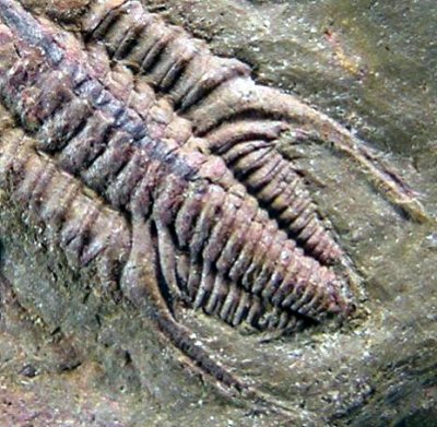
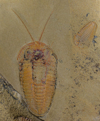
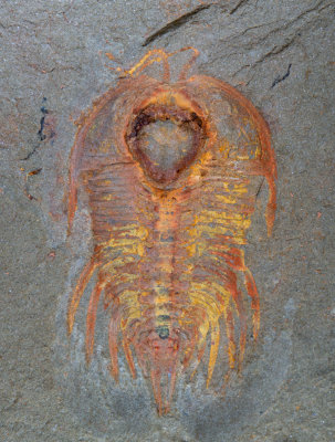
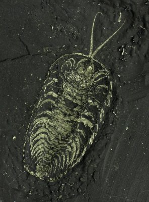
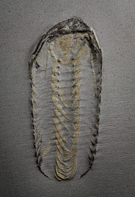
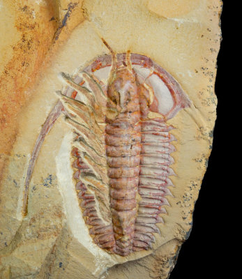
Eoredlichia intermedia, 5 cm long, Lower Cambrian, Chengjiang, China, partially dissected to show limbs and antennae.
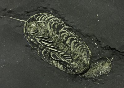
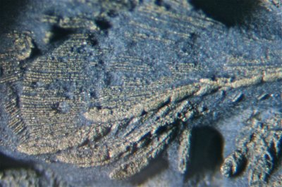
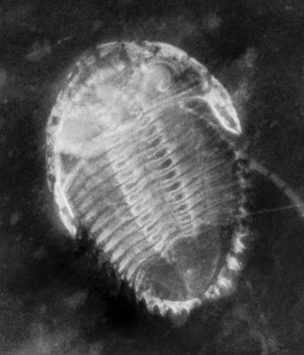
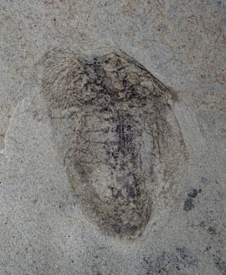
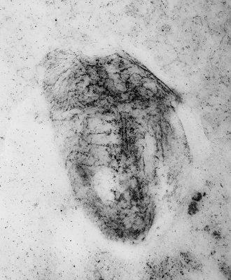
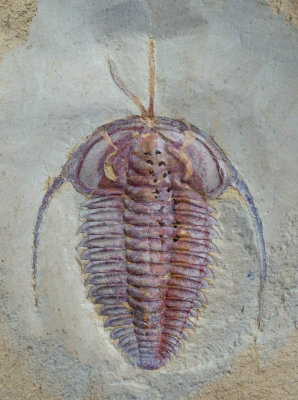
Eoredlichia intermedia (3 cm long), Lower Cambrian, Chengjiang, China, showing antennae and diverticulae.
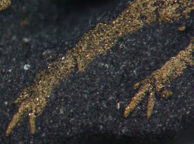
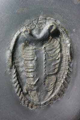
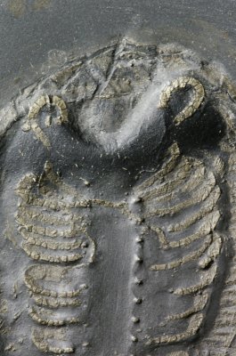
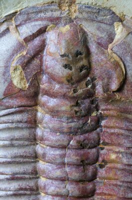
Detail of Eoredlichia showing paired gastric diverticulae. Glabella length is 8 mm.
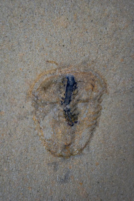
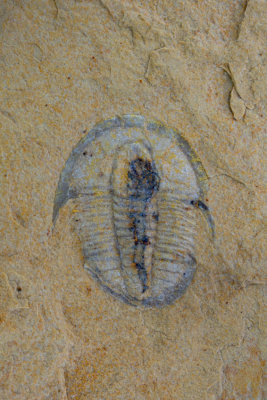
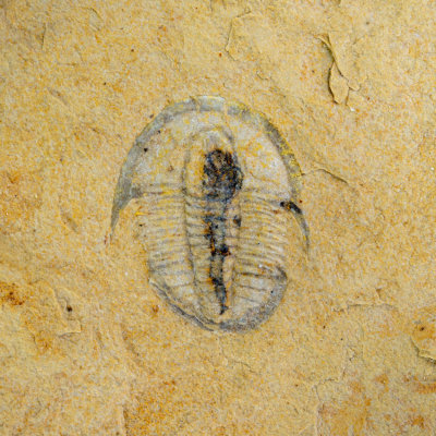
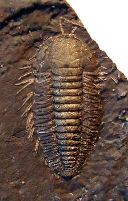
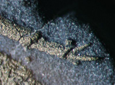
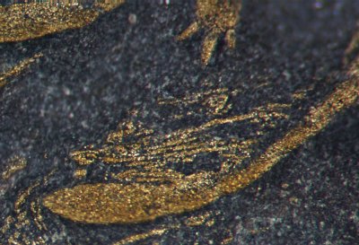
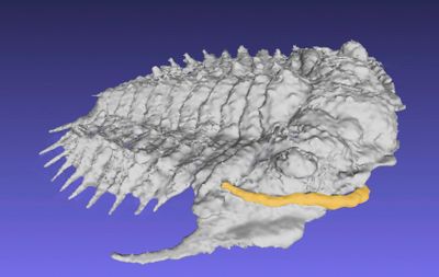
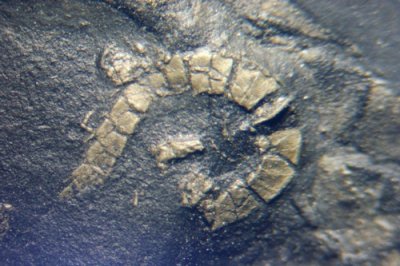
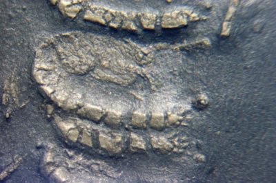
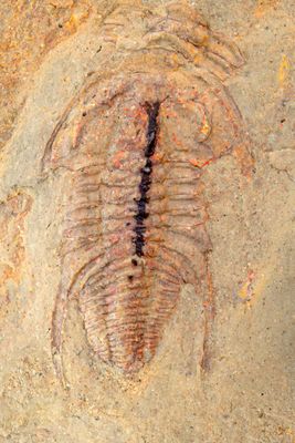
Balcoracania dailyi (12 mm long), Emu Bay Shale Fm, Lower Cambrian, S Australia, showing mineralized gut.
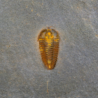
Eokochaspis piochensis, 13 mm with gut and antennae. Basal Middle Cambrian Comet Shale member, Pioche Formation, Nevada.
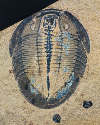
Meniscopsia beebei with mineralized gut and gastric caeca, 36 mm. Weeks Formation, M-U Cambrian, Utah, USA.
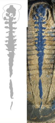
Detail of the Meniscopsia mineralized alimentary canal and caeca. The outline of the hypostome is shown.
Cedaria minor with preserved gut, 12 mm, Weeks Formation, M-U Cambrian, Utah, USA.
Bavarilla sp (4 cm) with spectacular complete antennae showing setal hairs, Lower Ordovician Fezouata Formation, Morocco
Bavarilla with antennae, Fezouata Formation, Zagora, Morocco
Anacheirurus sp., 32 mm long, with antennae, from the Fezouata of Morocco. Early pyrite has oxidised to colourful ochres.
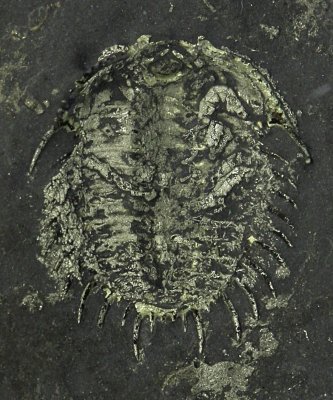
Triarthrus eatoni, 22mm showing limbs and antennae. Whetstone Gulf Formation, U Ordovician, Lewis Co, New York, USA.
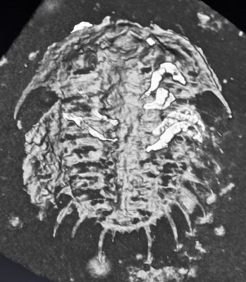
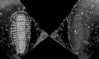
Dorsal and ventral views in micro CT scan of pyritized Triarthrus eatoni with limbs, 22 mm.
Detail showing eye and proximal antennae of a New York Triarthrus. Note dusting of pyrite micro crystals across the glabella.
Detail of Triarthrus antennae showing some fine setal hairs on proximal segments
Triarthrus eatoni, 49 mm showing limbs and antennae. Whetstone Gulf Formation, U Ordovician, Lewis Co, New York, USA.
Triarthrus eatoni, 45 mm showing limbs and antennae. Whetstone Gulf Formation, U Ordovician, Lewis Co, New York, USA.
Detail of Triarthrus claws
Detail of Triarthrus claw
Detail of Triarthrus exopods showing terminal paddles and filamentous lamellae.
Detail of an exopod terminal oar
Detail of an exopod terminal "oar"
Meadowtownella cf trentonensis, 9 mm, showing limbs and antennae. Lorraine Group, U Ordovician, Lewis County, New York.
Meadowtownella with limbs and antennae. CT image.
3D print, x7 of Meadowtownella with antennae marked in black
Meadowtownella extract from CT image showing swept back antenna
X-radiograph showing Rhenops sp, 45 mm long. Limbs are visible.
Rhenops sp, 45 mm ventral view, complete with limbs and antennae. Hunsrueckschiefer, L Devonian, Bundenbach, Germany.
Rhenops sp, 45 mm complete with limbs and antennae. Hunsrueckschiefer, L Devonian, Bundenbach, Germany.
Rhenops detail of antenna, like the Triarthrus showing setal hairs
Rhenops detail showing limbs with claws
Chotecops ferdinandi, 55 mm complete with limbs and antennae. Hunsrueckschiefer, L Devonian, Bundenbach, Germany. X-radiograph
Chotecops ferdinandi, 55 mm complete with limbs and antennae. Hunsrueckschiefer, L Devonian, Bundenbach, Germany.
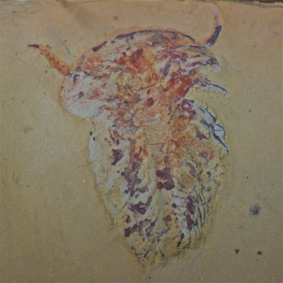
Naraoia spinosa, 15 mm long, showing antennae and limb preservation. Heilinpu Formation, Lower Cambrian, Chengjiang, China.
Naraoia, 2 cm showing alimentary canal, digestive diverticula and faint traces of limbs, Marjum Formation, Middle Cambrian, Utah
Naraoia, Marjum Formation, adjusted image
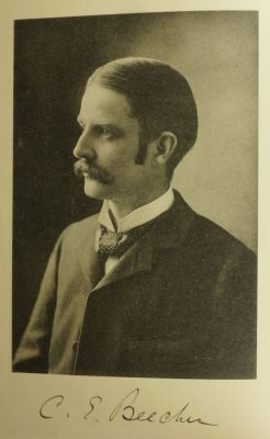
C.E. Beecher, frontispiece to Raymond (1920)
Beecher dedication in Raymond (1920)
Restoration of Olenoides serratus, Fig. 8 in Raymond (1920), Neolenus at the time.
Restoration of Olenoides serratus, Fig. 8 in Raymond (1920), "Neolenus" at the time.
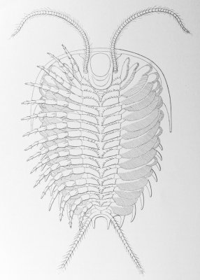
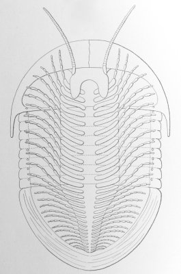
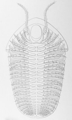
Restoration of Triarthrus, Fig. 10 in Raymond (1920)
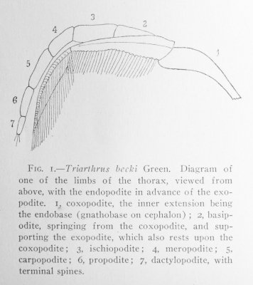
Reconstruction of Triarthrus biramous appendage by Raymond (1920).
Restoration of Cryptolithus, Fig. 20 in Raymond (1920)
Restoration of Isotelus gigas, Fig. 9 in Raymond (1920), based on Walcott's thin sections and a specimen from Ohio.
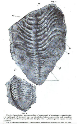
Mickleborough (1881)
Restoration of Calymene (now Flexicalymene) senaria, Fig. 16 in Raymond (1920).
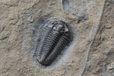
Flexicalymene senaria, 22 mm long. Trenton Group, Rust Formation, Spillway Member, Walcott-Rust Quarry, Russia, New York.
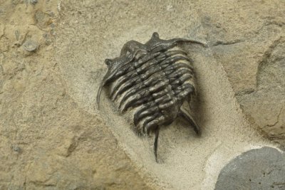
Ceraurus pleurexanthemus, partially enrolled, 25 mm. Trenton Group, Rust Formation, Spillway Member, Walcott-Rust Quarry, N York
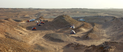
Camping on the Cambrian
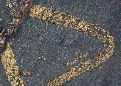
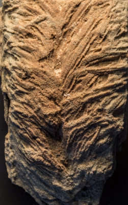
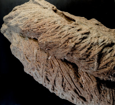
Cruziana omanicus, Cambrian, Huqf, Oman. Width of trace 4 cm.
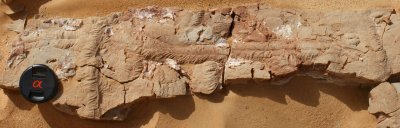
Cruziana omanicus, Cambrian, Huqf, Oman. Width of traces about 4 cm.
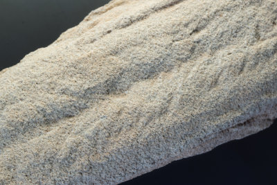
Cruziana semiplicata, Cambrian, Huqf, Oman. width 3 cm.
Trachomatichnites or galloping form of Diplichnites (Seilacher 2007 Plate 8).
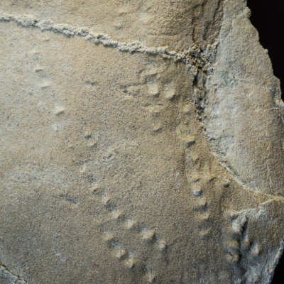
"Trachomatichnites" or "galloping" form of Diplichnites (Seilacher 2007 Plate 8).