


|
|
|
|
|
|
| Sherif Elwan | profile | all galleries >> OPHTHALMOLOGY LECTURES >> UNDERGRADUATE OPHTHALMOLOGY >> UNDERGRADUATE ATLAS OF OPHTHALMOLOGY | tree view | thumbnails | slideshow |
 Slide1.JPG |
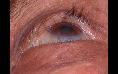
 1.Micro-cornea |
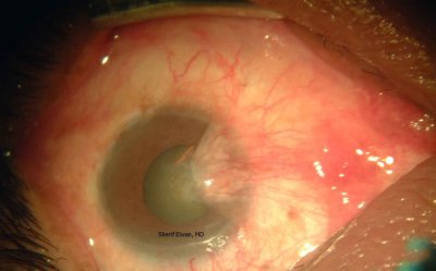
 2.Corneal Ulcer |
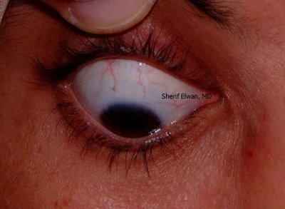
 3.Anterior Staphyloma |
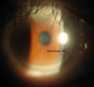
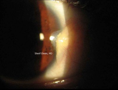
 4.Ateromatous Corneal Ulcer |
 5.PKP |
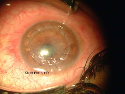
 6.PKP |
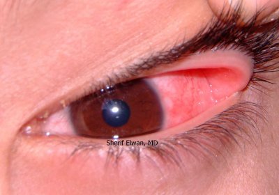
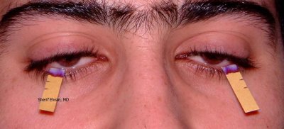
 7.Upper Lid Rubbing Lashes |
 8.Corneal Foreign Body |
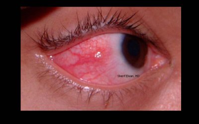
 9.Corneal Vascularization |
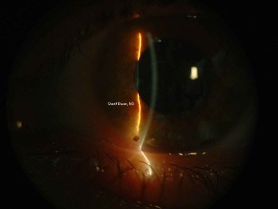
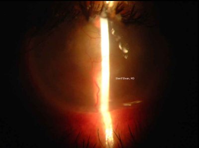
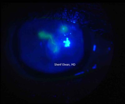 10.Dendritic Corneal Ulcer |
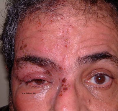 11.Herpes Zoster Ophthalmicus |
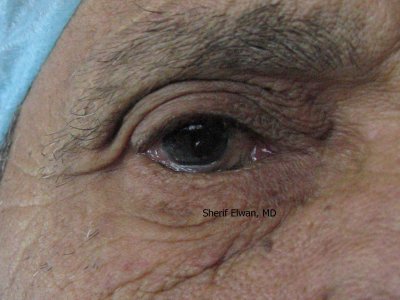
 12.Arcus Senilis |
 13.Central Corneal Scar |
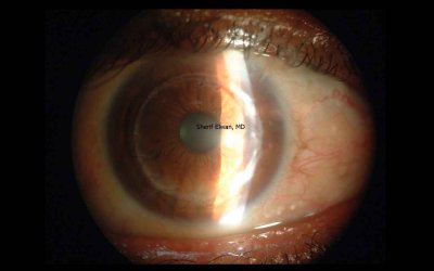
 14.Corneal Opacity |
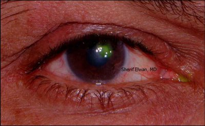
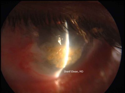
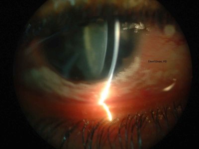
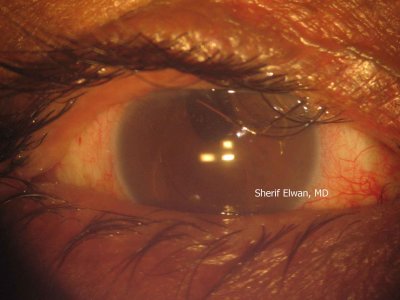
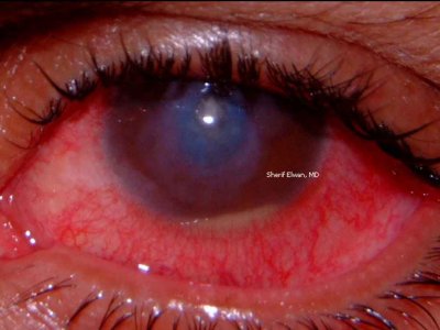 15.Corneal Ulcer with Hypopyon |
 16.Keratoconus: Munson's Sign |
 17.Pterygium |
 18.Advancing Pterygium |
 19.Bitots Spots |
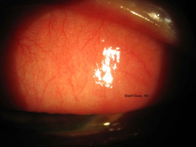
 20.Limbal Phlycten |
 21.Conjunctival Phlycten |
 22.Conjunctival Phlycten with Staphylococcal Blepharitis |
 23.Conjunctival Phlycten with Staphylococcal Blepharitis |
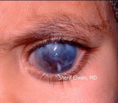
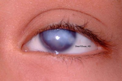
 24.Bilateral Primary Congenital Glaucoma: Buphthalmos |
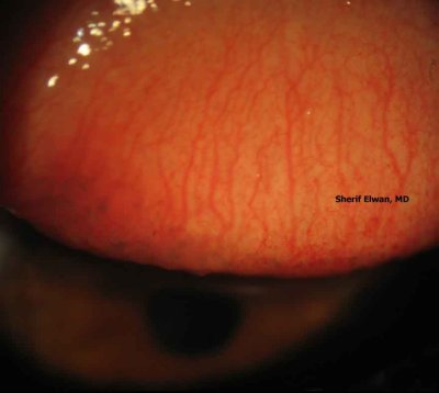
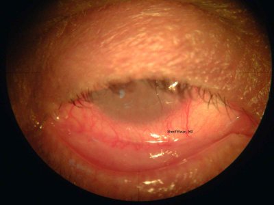
 25.Conjunctival Follicles |
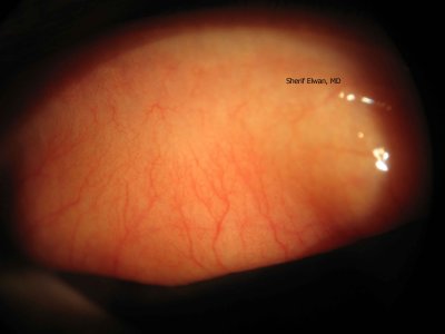
 26.Conjunctival Papillae |
 27.Arlt's Line |
 28.PTDs |
 29.Adult Acute Dacryo-cystitis |
 30.Child Acute Dacryo-cystitis |
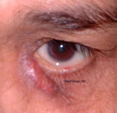
 31.Lacrimal Mucocele |
 32.Schirmer's Test |
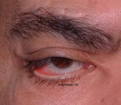
 33.Pre-septal cellulitis |
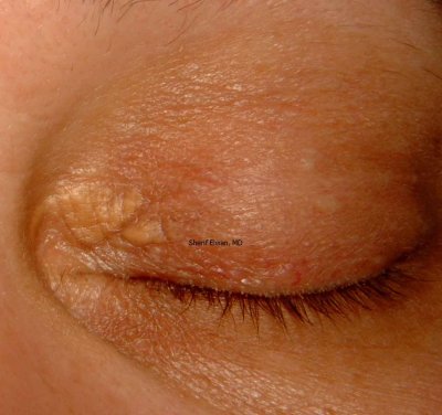
 34.Meibomian Gld Dysfunction |
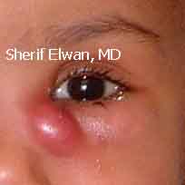
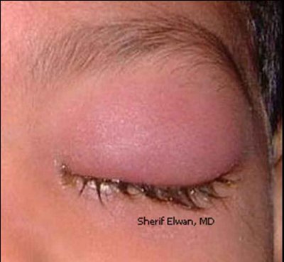
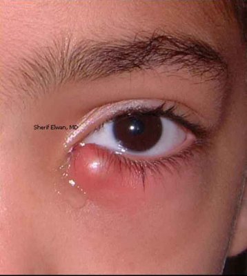
 35.Infected Upper Lid Chalazion |
 36.Retinoblastoma (Amourotic Cat's Eye) |
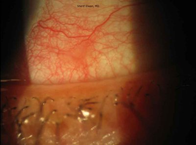
 37.Lower Lid Paralytic Ectropion: VIIth-nerve Palsy |
 38.Bilateral Congenital Ptosis |
 39.Lower Entropion Trichiasis |
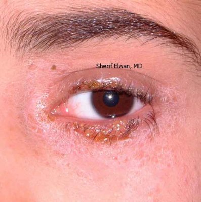 40. Staphylococcal Blepharitis |
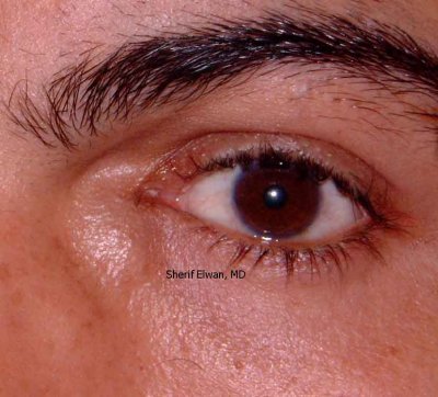
 41.Xanthelasma |
 42.Upper Entropion - Trichiasis |
 43.Staphylococcal Blepharitis |
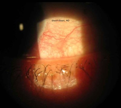
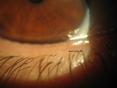
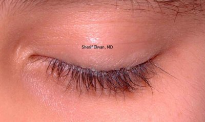
 44.Upper Multiple Rubbing Lashes |
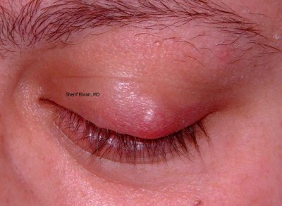
 45.Lower Infected Chalazion |
 46.Lateral Tarsorrhaphy |
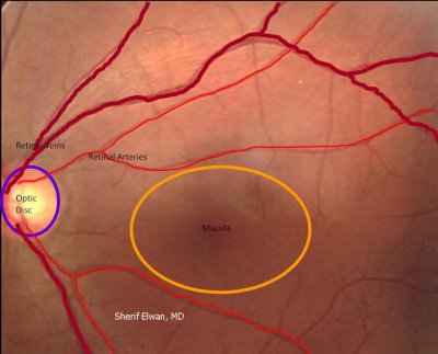
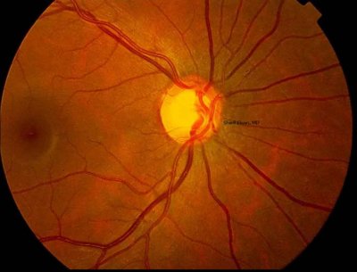
 47.Normal Fundus |
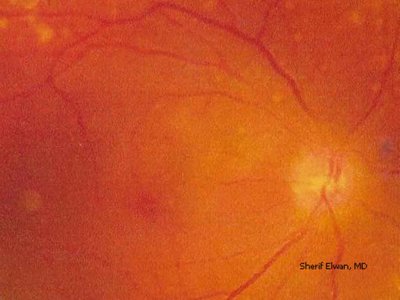
 48.Normal Fundus 2 |
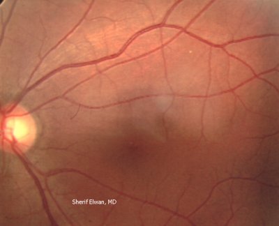
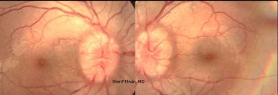
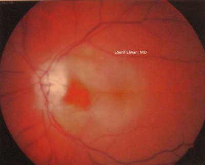
 49.Papillitis |
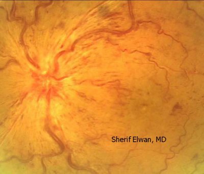
 50.Papilloedema |
 51.Central Retinal Vein Occlusion |
 52.Central Retinal Artery Occlusion |
 53.Arterial Emboli |
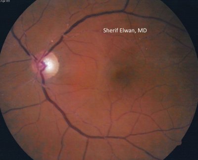
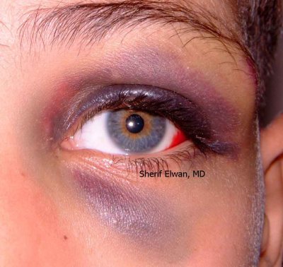
 54.Retinitis Pigmentosa |
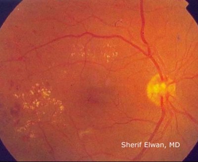
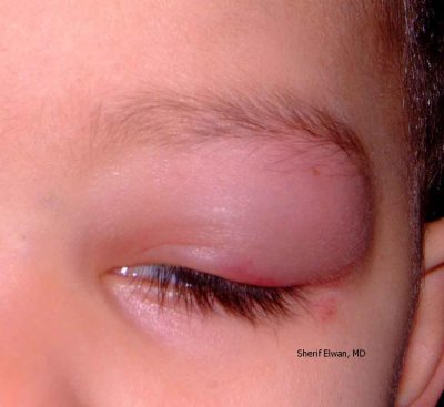
 55.Background Diabetic Maculopathy |
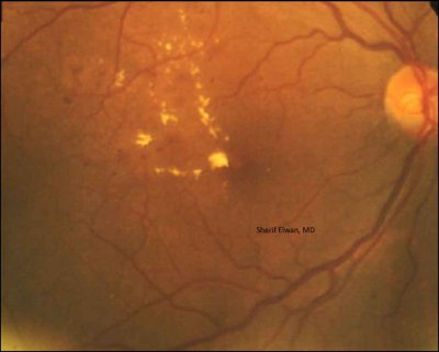
 56.Pre-proliferative Diabetic Retinopathy: Soft Exudates |
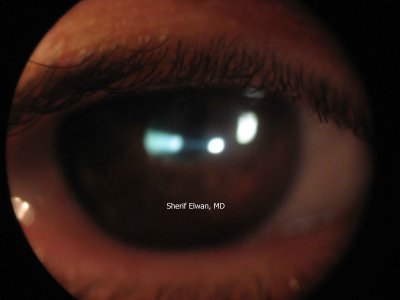
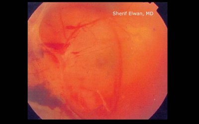 57.Pre-proliferative Diabetic Retinopathy: Pre-retinal Haemorrhage |
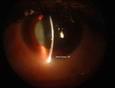
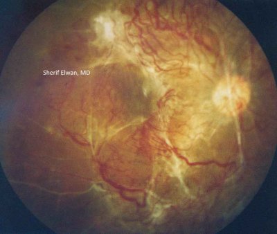 58.Proliferative Diabetic Retinopathy |
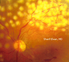 59.Recent Retinal Argon Laser Marks |
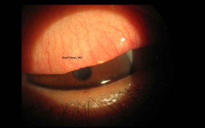
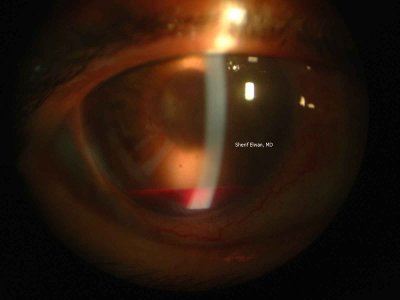 60.Hyphaema with a Level |
 61.Black Eye due to Blunt Trauma |
 62.Lid Haematoma due to Blunt Trauma |
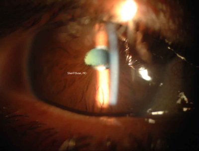
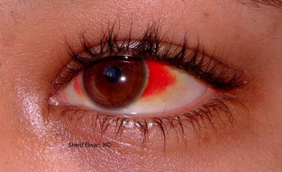 63.Subconjunctival Haemorrhage |
 64.Aqueous Flare |
 65.Keratitic Precipitates |
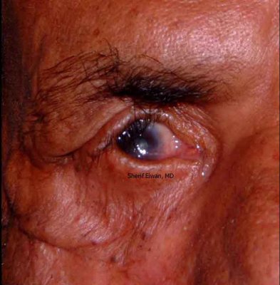
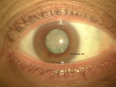
 66.Iris Neovascularization |
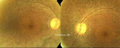
 67.Glaucomatous Disc Cupping: 0.6 |
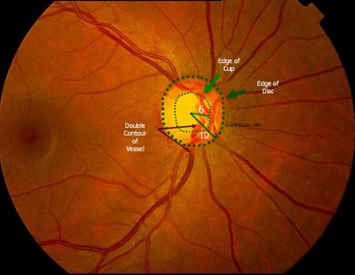 68.Glaucomatous Disc Cupping 0.6: Illustrated.JPG |
 69.Iris Neovascularization with Neovascular Membrane Extending Over Crystalline Lens |
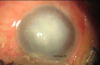
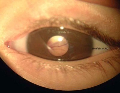
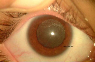
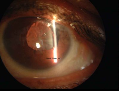
 70.Anterior Polar Cataract |
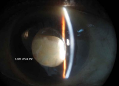
 71.Complicated Cataract, Posterior Synechiae, Festooned Pupil |
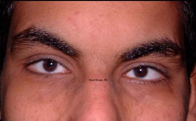
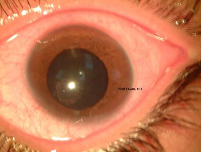
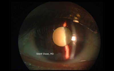
 72.Diabetic Cataract. |
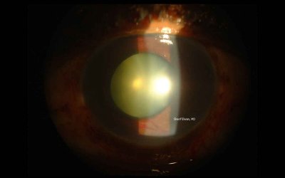
 73.Mature Senile Cataract |
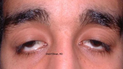
 74.Cataract with Pseudoexfoliation |
 75.Subluxated Mature Catactous Lens |
 76.Subluxated Clear Lens - Corneal Stitches |
 77.Posterior Cortical Cataract |
 78.Hypermature Morgagnian Cataract. |
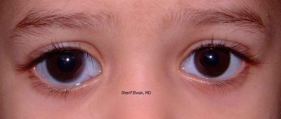
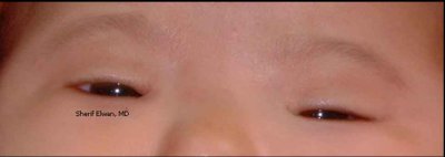 79.Apparent Convergent Squint: Medial Epicanthi |
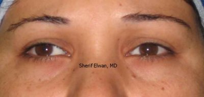
 80.Right Convergent Squint |
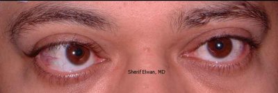
 81.Right Divergent Squint |
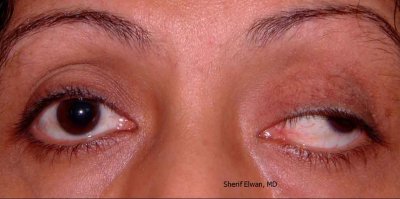 82.Left Oculomotor (III) Nerve Palsy: Ptosis & Divergent quint |
| comment | share |
| Mohamed | 19-Jul-2011 15:13 | |
| El Munibari | 30-Mar-2010 22:24 | |