


|
|
|
|
|
|
| lbliss | profile | all galleries >> weird knee injury | tree view | thumbnails | slideshow |
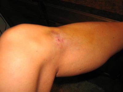
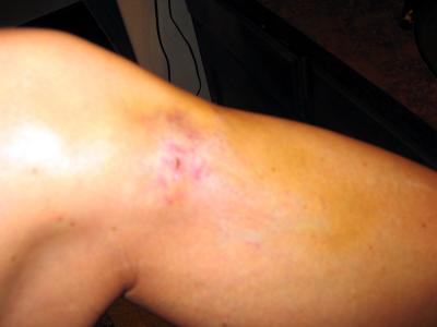
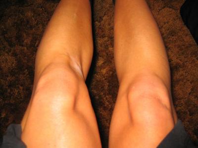 Subcutaneous tissue atrophy after a steroid injection into the left pes anserinus bursa in on Sept 4, 2004 |
 Skin, bone, tendons, vessels, hypopigmentation. No fat left. This is likely permanent. |
 the medial hamstring tendon insertions are clearly visible, as is the atrophy of the medial thigh and calf |
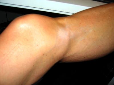
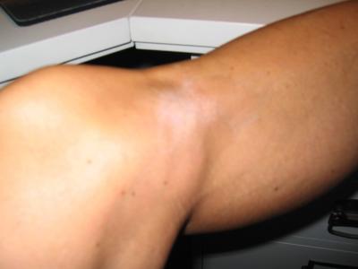 there are still steroid crystals visible (but not so clear here) under the skin in the center of the hypopigmented area |
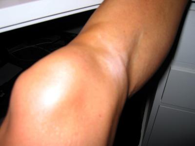
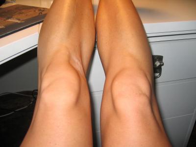 medial and lateral inferior pole atrophy makes the patellar tendon look prominent |
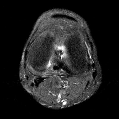 MRI axial left knee (1): thinning of the skin on the medial side and mild subcutaneous fluid |
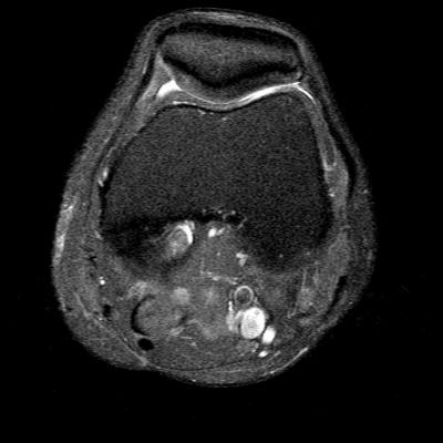 MRI axial left knee (2): Thin and incompetent medial patellofemoral ligament |
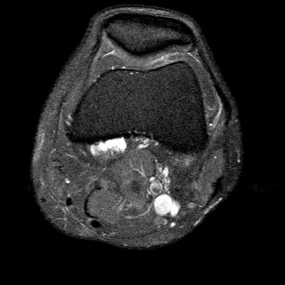 MRI axial left knee (3): the start of multilobulated perimeniscal cysts |
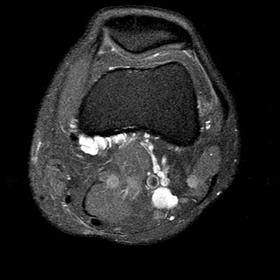 MRI axial left knee (4): the cysts grow & appear to be around the origin of the medial gastroc |
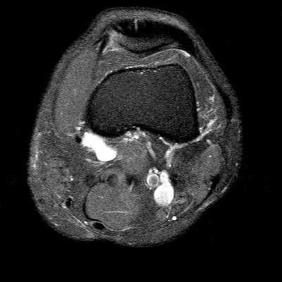 MRI axial left knee (5) |
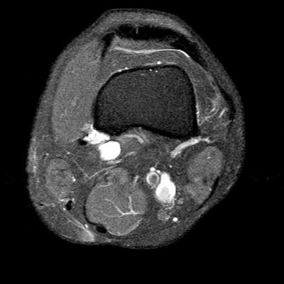 MRI axial left knee (6) |
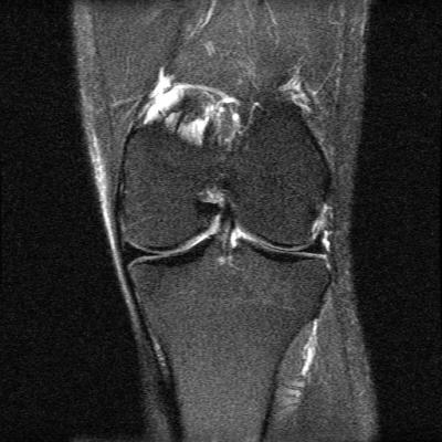 long view: medial subcutaneous edema and atrophy. Great looking menisci. |
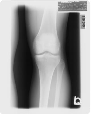 Normal AP plain film of the left knee: no joint space narrowing, no spurs - good as new. |
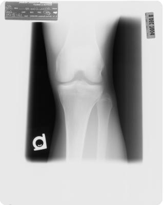 Normal weight-bearing plain film. |
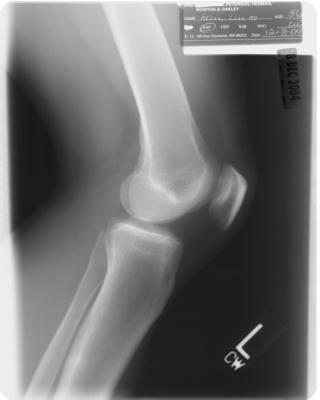 Normal lateral film |
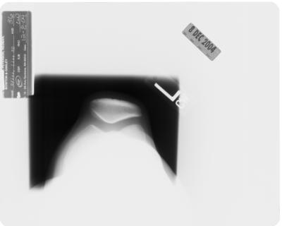 Normal sunrise view. Perfectly retained joint space. And the secret is.....Glucosamine! |
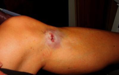 POD #0 s/p removal of 10 steroid crystals |
 A little Versed and Fentanyl enabled me to sit up and watch the procedure. |
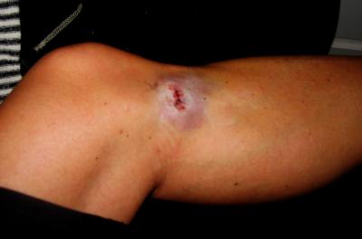
 POD #2. Healing. |
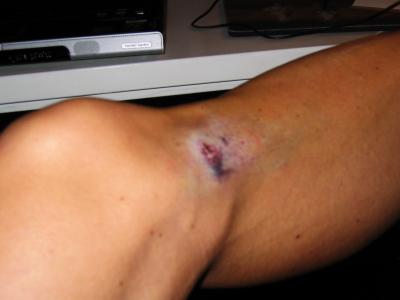
 POD #11. Sutures removed POD #8, steri's in place. PT continues.... |
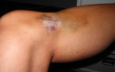
 2 weeks post-op |
 2 weeks post-op and getting back to normal |
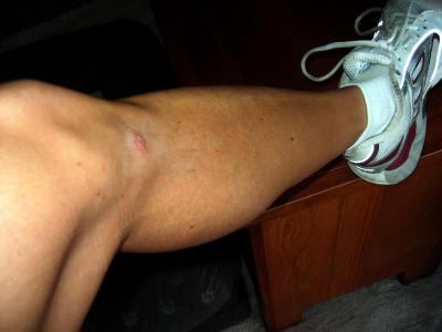 3 weeks post-op: note the running shoes! (but also the lingering medial thigh atrophy) |
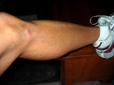 3 weeks post-op: I will try to run on the mountain tomorrow morning |
| comment | share |